Surgeon: Reza Razeghinejad, MD | Narrator: Lindsay Machen, MD | Year: 2019
PREOPERATIVE DIAGNOSIS:
Glaucoma, *** eyePOSTOPERATIVE DIAGNOSIS:
SamePROCEDURE PERFORMED:
Trabeculectomy with mitomycin-C, *** eyeATTENDING SURGEON:
***ASSISTANT SURGEON:
***ANESTHESIA:
MAC and topicalCOMPLICATIONS:
NonePrior to the date of surgery, the risks, benefits, and alternatives of the planned procedure were discussed with the patient, and informed consent was obtained. The patient was identified in the preoperative area by the attending physician, and the operative eye was marked. The patient was taken to the operative suite and given IV sedation and topical ocular anesthesia. The operative eye was prepped and draped in the usual sterile ophthalmic fashion. A lid speculum was inserted.
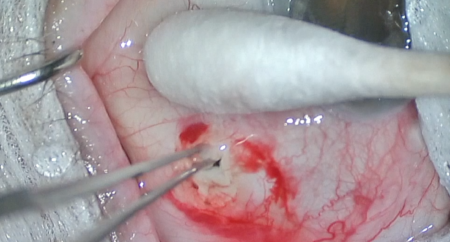
A 7-0 Vicryl traction suture was placed in partial-thickness superior cornea, and the eye was rotated inferiorly. A superior limbal conjunctival opening was made using Wescott scissors, then a 1:1 mixture of preservative-free 1% lidocaine and mitomycin-C (0.4 mg/mL) was injected into the subconjunctival space and spread using a blunt instrument. A peritomy was made superiorly, then blunt dissection was performed posteriorly with Westcott scissors. Gentle cautery was used to achieve hemostasis. A partial thickness scleral flap was created using a curved crescent blade.
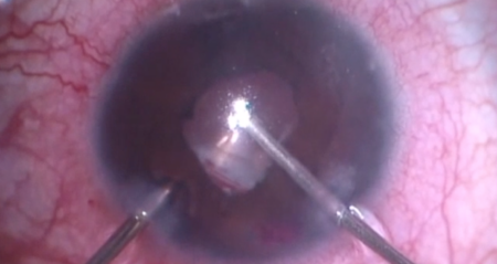
A SuperSharp blade was used to create a paracentesis through inferotemporal clear cornea. Viscoelastic was injected into the anterior chamber. The SuperSharp blade was then used to enter the anterior chamber at the base of the scleral flap. A Kelly punch was used to make an ostium. Vannas scissors and 0.12 forceps were used to create a surgical iridectomy. Two 10-0 nylon sutures were placed at the posterior edge of the scleral flap and adjusted to appropriate tension. Balanced salt solution was injected through the paracentesis to irrigate out the viscoelastic and test for flow across the scleral flap. After the flow was judged to be adequate, the nylon sutures were tied and the knots were buried. Next, 10-0 nylon suture was used to close the conjunctiva. The anterior chamber was refilled with balanced salt solution, and the bleb elevated easily.
The traction suture was removed. The corneal incision was hydrated then inspected with a Weck-cel sponge and found to be watertight with good intraocular pressure by finger tension. The speculum and drapes were removed, and antibiotic-steroid drops were placed in the eye. A shield was applied. The patient was then transported to the postoperative care unit in good condition.