Surgeon: Reza Razeghinejad, MD | Year: 2019
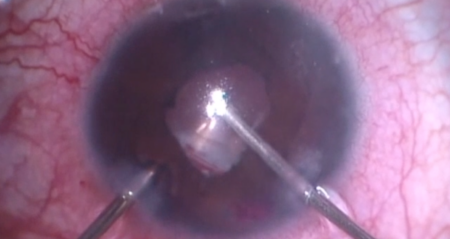
Dr. Razeghinejad performs a suprachoroidal effusion drainage. A conjunctival cut-down is created 5-6 mm posterior to the limbus. A triangular sclerotomy is made to access the suprachoroidal space, and serous fluid is drained. The anterior chamber is pressurized using viscoelastic. The sclerotomy flap is excised to allow the drainage to continue, then the conjunctiva is closed.
PREOPERATIVE DIAGNOSIS:
Hypotony and choroidal effusion, *** eyePOSTOPERATIVE DIAGNOSIS:
SamePROCEDURE PERFORMED:
Choroidal effusion drainage, *** eyeATTENDING SURGEON:
***ASSISTANT SURGEON:
***ANESTHESIA:
MAC and topicalCOMPLICATIONS:
NonePrior to the date of surgery, the risks, benefits, and alternatives of the planned procedure were discussed with the patient, and informed consent was obtained. The patient was identified in the preoperative area by the attending physician, and the operative eye was marked. The patient was taken to the operative suite and given IV sedation and topical ocular anesthesia. The operative eye was prepped and draped in the usual sterile ophthalmic fashion. A lid speculum was inserted.
Indirect ophthalmoscopy was performed and confirmed large choroidal effusions. A paracentesis was made into clear cornea. A conjunctival cut-down was performed over the choroidal effusion using Wescott scissors. A sclerotomy flap was created with a #69 blade until the suprachoroidal space was accessed and fluid was drained. The conjunctiva was then closed with 8-0 Vicryl sutures.
The anterior chamber maintainer was removed. The corneal incision was hydrated with balanced salt solution then inspected with a Weck-cel sponge and found to be watertight. There was a diffuse bleb, and the eye had good intraocular pressure by finger tension. The speculum and drapes were removed, and antibiotic-steroid drops were placed in the eye. A patch and shield were applied. The patient was then transported to the postoperative care unit in good condition.