Surgeon: Aakriti Garg Shukla, MD | Narrator: Lindsay Machen, MD | Year: 2019
PREOPERATIVE DIAGNOSIS:
Glaucoma, *** eyePOSTOPERATIVE DIAGNOSIS:
SamePROCEDURE PERFORMED:
Endocyclophotocoagulation, *** eyeATTENDING SURGEON:
***ASSISTANT SURGEON:
***ANESTHESIA:
MAC, topical, and localCOMPLICATIONS:
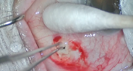
NonePrior to the date of surgery, the risks, benefits, and alternatives of the planned procedure were discussed with the patient, and informed consent was obtained. The patient was identified in the preoperative area by the attending physician, and the operative eye was marked. The patient was taken to the operative suite and given IV sedation and topical ocular anesthesia. The operative eye was prepped and draped in the usual sterile ophthalmic fashion. A lid speculum was inserted.
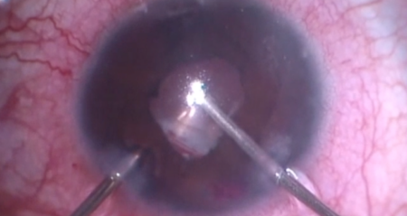
A sub-Tenon’s block was given using a 50:50 mixture of 4% lidocaine and 0.75% Marcaine. The endolaser was set to a power of *** with continuous duration. A 2.4 mm keratome was used to make a temporal clear corneal incision, then the nasal sulcus was inflated with viscoelastic. The endolaser was used to heat the nasal 180 degrees of ciliary processes until each whitened and shrunk. This process was repeated for the nasal ciliary processes. A Simcoe canula was used to remove the viscoelastic.
The corneal incisions were hydrated with balanced salt solution then inspected with a Weck-cel sponge and found to be watertight with good intraocular pressure by finger tension. Decadron was injected into the subconjunctival space. The speculum and drapes were removed, and antibiotic-steroid ointment was placed on the eye. A patch and shield were applied. The patient was then transported to the postoperative care unit in good condition.