Surgeon: Mark F. Pyfer, MD, FACS | Year: 2019
Dr. Pyfer uses the OMNI device to perform an ab-interno canaloplasty and viscodilation of the collector channels.
PREOPERATIVE DIAGNOSIS:
Glaucoma, *** eyePOSTOPERATIVE DIAGNOSIS:
SamePROCEDURE PERFORMED:
OMNI canaloplasty, *** eyeATTENDING SURGEON:
***ASSISTANT SURGEON:
***DEVICE:
OMNI Surgical System, serial number ***ANESTHESIA:
MAC and topicalCOMPLICATIONS:
NonePrior to the date of surgery, the risks, benefits, and alternatives of the planned procedure had been discussed with the patient, and informed consent had been obtained. The patient was identified in the preoperative area by the attending physician, and the operative eye was marked. The patient was transported to the operative suite and given IV sedation and topical ocular anesthesia. The operative eye was prepped and draped in the usual sterile ophthalmic fashion. A lid speculum was inserted to provide adequate exposure.
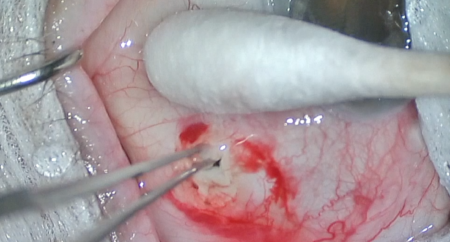
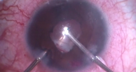
A paracentesis was made into clear cornea. Epi-Shugarcaine and viscoelastic were injected into the anterior chamber. A clear corneal incision was made using a keratome. The patient’s head was then rotated 35 degrees away from the surgeon, and the operating microscope was rotated 35 degrees toward the surgeon. Additional viscoelastic was injected into the anterior chamber to inflate the nasal angle and onto the cornea to serve as a coupling agent. A direct gonioprism was used to visualize the nasal angle. The trabecular meshwork was easily identified. The OMNI device was primed then used to catheterize and viscodilate Schlemm’s canal 180 degrees in both directions. [Next, the OMNI cannula was readvanced through Schlemm’s canal, then the handpiece was retracted through the paracentesis to perform a 180-degree goniotomy. Adequate incision of the trabecular meshwork was achieved with mild bleeding.]
The patient’s head and the operating microscope were rotated back into neutral positions. All remaining viscoelastic was irrigated using balanced salt solution. The corneal incisions were hydrated with balanced salt solution then inspected with a Weck-cel sponge and found to be watertight with good intraocular pressure by finger tension. The speculum and drapes were removed, and antibiotic-steroid drops were placed into the eye. The patient was then transported to the postoperative care unit in good condition.