Surgeon: Ravi Pandit, MD, MPH | Year: 2019
Dr. Pandit performs a pars plana vitrectomy and epiretinal membrane peel for macular pucker. First, 23-gauge trochars are placed, and a core vitrectomy is performed. Next, indocyanine green is instilled to stain the internal limiting membrane (ILM). Under high magnification, an ILM flap is created, then the ILM is peeled circumferentially around the macula. Care is taken to minimize the instruments crossing the fovea to avoid phototoxicity or mechanical trauma. Finally, 360-degree scleral depression is performed to identify any retinal breaks, and an air fill is performed.
PREOPERATIVE DIAGNOSIS:
Macular pucker, *** eyePOSTOPERATIVE DIAGNOSIS:
SamePROCEDURE PERFORMED:
1. Pars plana vitrectomy, *** eyeATTENDING SURGEON:
***ASSISTANT SURGEON:
***ANESTHESIA:
MAC, topical, and peribulbar blockCOMPLICATIONS:
NonePrior to the date of surgery, the risks, benefits, and alternatives of the planned procedure were discussed with the patient, and informed consent was obtained. The patient was identified in the preoperative area by the attending physician, and the operative eye was marked. The patient was taken to the operative suite and given IV sedation and topical ocular anesthesia. The operative eye was prepped and draped in the usual sterile ophthalmic fashion. A lid speculum was inserted. A sub-Tenon block consisting of 50:50 lidocaine/marcaine was delivered inferonasally.
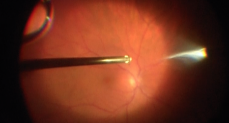
A 23-gauge infusion cannula was inserted in the inferotemporal quadrant using the self-retaining port. Placement in the vitreous cavity was confirmed using direct visualization, then infusion was initiated. Self-retaining 23-gauge cannulas were inserted in the superonasal and superotemporal quadrants. The light pipe and vitrector were inserted into the vitreous cavity. Visualization confirmed an epiretinal membrane and a posterior vitreous detachment. A core vitrectomy was performed, followed by a peripheral shave vitrectomy.
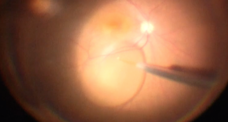
Indocyanine green dye was injected to visualize the internal limiting membrane. A flat contact lens was placed. ILM forceps were used to peel the internal limiting membrane and epiretinal membrane atraumatically off the macula from arcade to arcade. Widefield viewing was restored, then the vitrector was used to aspirate any remaining membrane fragments.
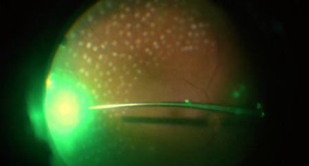
The peripheral retina was meticulously inspected with scleral depression, and there were no retinal breaks present. A partial air-fluid exchange was performed. The ports were removed, and the sclerostomies were inspected and found to be watertight. The eye was physiologic pressure by finger tension. Subconjunctival antibiotic was injected into the inferior fornix. The speculum and drapes were removed, antibiotic-steroid drops were placed in the eye, and a shield was placed over the eye. The patient was then transported to the recovery unit in stable condition.