Surgeon: Aakriti Garg Shukla, MD | Narrator: Lindsay Machen, MD | Year: 2019
PREOPERATIVE DIAGNOSIS:
Cyclodialysis cleft, *** eyePOSTOPERATIVE DIAGNOSIS:
SamePROCEDURE PERFORMED:
Cyclodialysis cleft repair, *** eyeATTENDING SURGEON:
***ASSISTANT SURGEON:
***ANESTHESIA:
MAC, topical, and localCOMPLICATIONS:
NonePrior to the date of surgery, the risks, benefits, and alternatives of the planned procedure were discussed with the patient, and informed consent was obtained. The patient was identified in the preoperative area by the attending physician, and the operative eye was marked. The patient was taken to the operative suite and given IV sedation and topical ocular anesthesia. A peribulbar block was administered. The operative eye was then prepped and draped in the usual sterile ophthalmic fashion. A lid speculum was inserted.
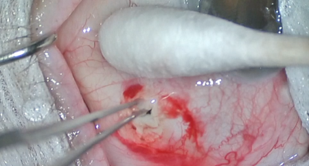
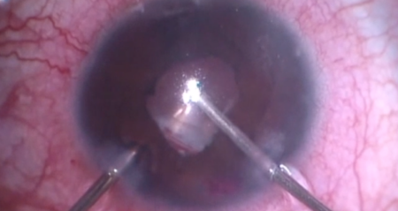
The eye was examined using a gonioprism, and the cyclodialysis cleft was identified. The conjunctiva was incised at the limbus to create a fornix-based conjunctival flap. Light cautery was used to achieve hemostasis. A 4-mm, half-thickness scleral flap was raised over the cyclodialysis cleft. The scleral spur was identified. The anterior chamber was entered through the scleral bed over the scleral spur. Two posterior incisions were made at the lateral edges of the scleral bed so that an internal scleral flap could be raised, allowing direct visualization of the cyclodialysis cleft. Viscoelastic was injected into the anterior chamber. Interrupted 10-0 nylon sutures were placed with the initial bites underneath the flap in the anterior lip of the incision, then in the ciliary body, and then through the posterior edge of the internal flap. These sutures were then tied, which pulled the ciliary body back to the scleral wall.
The area was re-examined with the gonioprism, and it was noted that the cleft had been closed. The scleral flap was reapproximated with interrupted 10-0 nylon sutures. The anterior chamber was reformed with balanced salt solution to physiologic pressure. The conjunctival flap was reapproximated to the limbus with interrupted 8-0 Vicryl sutures.
The speculum and drapes were removed, and antibiotic-steroid drops were placed in the eye. A patch and shield were applied. The patient was then transported to the postoperative care unit in good condition.