Surgeon: Aakriti Garg Shukla, MD | Year: 2019
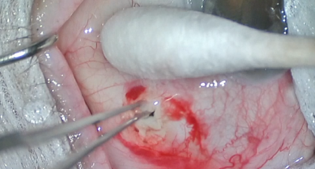
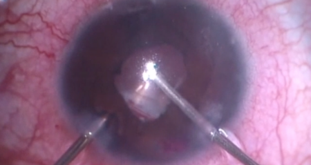
Dr. Shukla implants an Ahmed tube shunt in a recently-ruptured globe. First, the rupture site is explored, and a scleral patch graft is placed over exposed uvea. Next, a superotemporal peritomy is made and a sub-Tenon’s block given. The Ahmed valve is primed, then the plate is placed 9 mm posterior to the limbus and secured to the sclera. The tube is trimmed, and a 23-gauge needle is tunneled into the anterior chamber. The tube is fed into the anterior chamber, secured to the sclera using a 9-0 Nylon figure-of-eight suture, then covered with a split-thickness corneal patch graft. Finally, the conjunctiva is closed with Vicryl sutures.
PREOPERATIVE DIAGNOSIS:
Glaucoma, *** eyePOSTOPERATIVE DIAGNOSIS:
SamePROCEDURE PERFORMED:
Ahmed Glaucoma Valve implantation, *** eyeATTENDING SURGEON:
***ASSISTANT SURGEON:
***IMPLANTS:
1. Ahmed Glaucoma Valve, serial number ***ANESTHESIA:
MAC, topical, and localCOMPLICATIONS:
NonePrior to the date of surgery, the risks, benefits, and alternatives of the planned procedure had been discussed with the patient, and informed consent had been obtained. The patient was identified in the preoperative area by the attending physician, and the operative eye was marked. The patient was transported to the operative suite and given IV sedation and topical ocular anesthesia. The operative eye was prepped and draped in the usual sterile ophthalmic fashion. A lid speculum was inserted to provide adequate exposure.
A 7-0 Vicryl traction suture was placed in partial thickness superior cornea, and the eye was rotated infranasally. A superior limbal conjunctival opening was made using Wescott scissors. Bupivacaine was injected into the subconjunctival space and spread using a blunt instrument. A peritomy was made temporally through superiorly with radial relaxing incisions. Blunt dissection was performed with Westcott scissors to expand the pocket underlying conjunctiva and Tenon’s. A peribulbar block was given using bupivacaine. Gentle cautery was performed to achieve hemostasis at the intended site of tube entry.
The Ahmed implant was primed with balanced salt solution then placed into the supratemporal pocket. Two 8-0 nylon sutures were passed through partial thickness sclera 8 mm posterior to the limbus, threaded through the Ahmed plate eyelets, then tied. The plate was examined and found to be positioned well with no tendency to move forward. Westcott scissors were then used to cut the tube to an appropriate length in a beveled fashion.
Next, a paracentesis was made in clear cornea, then the anterior chamber was filled with viscoelastic. A 23-gauge bent needle was used to create a tunneled sclerostomy that entered the anterior chamber above the iris plane but deep to the corneal endothelium. The tube was inserted through this tract, positioned appropriately in the anterior chamber, then secured to the sclera with a 10-0 nylon suture. The Tutoplast processed pericardial patch graft was secured over the exposed tube with 7-0 Vicryl suture. The conjunctiva was advanced using smooth forceps and secured at the limbus with 7-0 Vicryl interrupted and running sutures.
The traction suture was removed. The corneal incision was hydrated with balanced salt solution then inspected with a Weck-cel sponge and found to be watertight with good intraocular pressure by finger tension. The tube was inspected and found to be in good position with respect to the cornea, iris, and lens. The speculum and drapes were removed, and antibiotic-steroid drops were placed in the eye. A patch and shield were applied. The patient was then transported to the postoperative care unit in good condition.