Surgeon: Brandon Ayres, MD | Year: 2016
Dr. Ayres removes a subluxed lens in a patient with Marfan syndrome and places a toric IOL with the assistance of the Catalys femtosecond laser.
PREOPERATIVE DIAGNOSES:
1. Cataract, *** eyePOSTOPERATIVE DIAGNOSES:
SamePROCEDURES PERFORMED:
1. Corneal astigmatism reduction using the femtosecond laser, *** eyeIMPLANT:
*** *** diopters, serial number ***, aim ***ATTENDING SURGEON:
***ASSISTANT SURGEON:
***ANESTHESIA:
MAC and topicalCOMPLICATIONS:
NonePrior to the date of surgery, the risks, benefits, and alternatives of the planned procedure were discussed with the patient, and informed consent was obtained. The patient was identified in the preoperative area by the attending physician, and the operative eye was marked. The patient was taken to the laser suite, and the operative eye was marked in the 3:00, 6:00, and 9:00 positions. The femtosecond laser was used to create the capsulotomy, fragment the lens, and create limbal relaxing incisions if indicated. The patient was then taken to the operative suite and given IV sedation and topical ocular anesthesia. The operative eye was prepped and draped in the usual sterile ophthalmic fashion. A lid speculum was inserted.
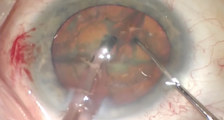
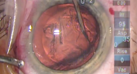
A paracentesis was made into clear cornea. Epi-Shugarcaine and viscoelastic were injected into the anterior chamber. A clear corneal incision was made using a keratome. The capsulorrhexis was removed, confirming a continuous capsulotomy without tears or rents. Hydrodissection was performed using balanced salt solution on a blunt tipped cannula. A phacoemulsification handpiece was used to disassemble and remove the lens nucleus. An irrigation/aspiration handpiece was used to remove the remaining cortical material and polish the capsule.
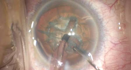
The anterior chamber and capsular bag were filled with viscoelastic. The intraocular lens was placed into the bag and dialed into position. All remaining viscoelastic was removed using the irrigation/aspiration handpiece. The corneal incisions were hydrated with balanced salt solution then inspected with a Weck-cel sponge and found to be watertight with good intraocular pressure by finger tension. The lens was inspected and found to be centered in the capsular bag.
The speculum and drapes were removed, and antibiotic-steroid drops were placed into the eye. The patient was then transported to the postoperative care unit in good condition.